

Cells are the structural and functional unit of all living organisms, and are described as "building block of life". As mentioned earlier, Cell was first discovered by Robert Hooke in 1665 in cork tissue.
The word Cell comes from the Latin cellula, a small room. Loewy and Sickevitz defined Cell as “a unit of biological activity de-limited by a semi-permeable membrane and capable of self-reproduction in a medium free of other living system”.
Depending on the number of cells, organisms are divided into Unicellular and Multicellular. While Bacteria, Blue and Green Algae and Protozoa are unicellular, other organisms such as plants and animals are multicellular. Humans have an estimated 100 trillion or 1014 cells; a typical cell size is 10 µm, a typical cell mass 1 nanogram). The largest known cell is an Ostrich egg.
The cell theory, first developed by German scientists Schleiden (1838) and Schwann (1839), states that all organisms are composed of one or more cells. Rudolf Virchow (1858) developed the idea of generation of cells by stating, “Omnis cellula e cellua”, which means cells arise from preexisting cells by cell division.
All living systems including both plant and animal are made up of cells and depending on the level of cellular organization, are of two major types: prokaryotic and eukaryotic.
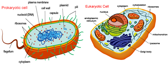
Comparison of Prokaryotic and Eukaryotic Cells
Prokaryotic cell
| | Prokaryotes | Eukaryotes |
| Typical organisms | Bacteria, Blue green algae, PPLOs (Pleuropneumonia like organisms) | Protists, Fungi, Plants, Animals |
| Typical size | ~ 1-10 µm | ~ 10-100 µm (sperm cells, apart from the tail, are smaller) |
| Organization | Usually single cells. | Single cells, colonies, higher multicellular organisms with specialized cells. |
| Type of nucleus | nucleoid region | True nucleus with double membrane. |
| DNA | Single Covalently Closed Circular DNA molecule. (usually) | Linear and multiple molecules (chromosomes). |
| Histones | Absent but histone like proteins are present. | Present |
| Compartmentalisation | Lacks well defined cytoplasmic organelles | Posses well defined cytoplasmic organelles like endosplasmic reticulum, golgi bodies, lysosomes, chloroplast, mitochondria. |
| RNA-/protein-synthesis | Transcription and Translation are coupled in cytoplasm | Transcription occurs in the nucleus. Translation occurs in cytoplasm |
| Ribosomes | 60S type (50S+30S) | 70S type (60S+40S) |
| Cytoplasmatic structure | Very few structures | Highly structured by endomembranes and a cytoskeleton |
| Cell movement | Flagella made of flagellin | Flagella and cilia made of tubulin |
| Respiration | Mesosomes support respiration. | Mitochondria support respiration. |
| Mitochondria | None | One to several dozen (though some lack mitochondria) |
| Pigments | Pigments are distributed throughout the | Pigments are present in plastids |
| Chloroplasts | Absent | In algae and plants |
| Cell division | Binaryfission (simple division) | Mitosis (fission or budding) |
Prokaryotic cells are relatively small (1–5mm in diameter) and simple. Single celled microorganisms or bacteria are fall into this category. These organisms lack a well defined nucleus (or karyon), having only a central area in the cell cytosol where the genetic material, the deoxyribonucleic acid (DNA), is found.
A Prokaryotic cell is characterized by the presence of a single, circular covalently closed DNA molecule (an exception is – that of the bacterium Borrelia burgdorferi causing Lyme disease contains linear chromosomes). However, in addition to the single chromosome, Prokaryotes carry extrachromosomal DNA elements called plasmids. Plasmids are usually circular and carry additional functions, such as antibiotic resistance. In fact the term Nucleoid is used to differentiate it from Nucleus. DNA is relatively naked, without associated histone protein. However histone like proteins like Hu and polyamines like spermine and spermidine are bound to the DNA. As these cells possess no distinct membrane-bound nucleus, they are Prokaryotic.
Eukaryotic (with a karyon) cells, in contrast, are the constituents of all multicellular organisms. They possess a distinct nucleus, bounded by a double nuclear envelope, wherein lies the genetic material in the form of the DNA packaged as chromosomes. This DNA is combined with histone protein to make up a nucleoprotein complex called chromatin or chromosomes which are linear. Such cells are much larger than prokaryotic cells, ranging in diameter from 10mm up to many millimetres (of which egg cells are the largest), although averaging about 30–60mm in diameter.
Differences in the cell size can be justified as prokaryotic cells lack internal compartmentalization (organelles) and on the other hand Eukaryotic cells have extensive subcellular compartments of different kinds. Biological processes like respiration, protein synthesis…etc are performed by various structures present in the cytoplasm.
In order to localize and separate these cellular events from one another, different compartments are generated with the help of membranes. Intracellular membrane bound compartments are termed as the organelles. Cells include organelles like endoplasmic reticulum, Golgi apparatus, mitochondria, chloroplasts and lysosomes in Eukaryotes in addition to an internal cytoskeleton.
The prokaryotic cell type is sufficiently small allowing for simple diffusion mediating efficient transport and exchange of materials across the plasma (cell) membrane. Some plasma membrane is double-membraned and also possess an outer cell wall, which provides support and strength to maintain the integrity of the cell during environmental stress. The cells contain a cytosol, or ground cytoplasm, which contains all the enzymes (catalysts) and building blocks (metabolites) to synthesize cell components. Internal compartmentalization is absent.
Comparison of structures between Animal and Plant Cells
|
PLANT CELL |
ANIMAL CELL |
|
Plant cell has a rigid wall on the outside. |
The cell wall is absent. |
| Usually larger in size. | Comparatively smaller in size. |
| Cannot change its shape. | Can often change its shape. |
| Plastids are found. | Plastids are usually absent. |
| Possess chlorophyll containing plastids called chloroplasts. | Chloroplasts are absent. |
| A mature plant cell contains a large central vacuole. | Vacuoles are numerous and very small. |
| Nucleus lies on one side in the peripheral Cytoplasm. | Nucleus usually lies in the centre. |
| Mitochondria are comparatively fewer. | Mitochondria are generally more numerous. |
| Cristae are tubular in plant mitochondria. | Cristae are plate like in animal mitochondria. |
| Plant cells do not burst if placed in hypotonic solution due to presence of cell wall. | Animal cells burst if placed in hypotonic solution unless and until it posses contractile vacuole. |
| Centrioles are usually absent in lower plants. | Centrioles are found in animal cell. |
| Spindle fibres formed during nuclear division are anastral. | Spindle fibres formed during nuclear division are amphiastral. |
| Golgi apparatus consists of a number of distinct / unconnected units called dictyosomes. | Golgi apparatus is either localized or consists a well connected single complex. |
| Cytoskeleton does not con tain intermediate fibres. | Cytoskeleton contains intermediate fibres. |
| Lysosomes are rare and their activity is performed by specialized vacuoles. | Typical lysosomes occur in animal cell. |
| Glyoxysomes may be present. | Glyoxysomes are absent. |
| Crystals of inorganic substances may occur inside the cells. | Crystals usually do not occur in animal cells. |
| Reserve food is generally starch and fat. | Reserve food is usually glycogen and fat. |
| A tissue fluid does not bathe the individual cells. | A tissue fluid contain in a NaCl bathes the cells. |
| Adjacent cells may be connected through plasmadesmata. | Adjacent cells are connected through a number of junctions. |
The Nucleus
The nucleus was first discovered by Scottish Botanist Robert Brown in 1831. While studying orchids microscopically, Brown characterized an opaque area, in the cells of the epidermis, which he called the areola or nucleus
Structure of Nucleus
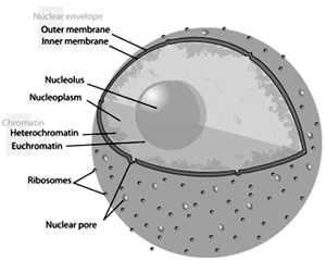
The outer nuclear membrane is continuous with the membrane of the rough endoplasmic reticulum, and is similarly studded with ribosomes. The space between the membranes is called the perinuclear space and is continuous with the lumen of the rough endoplasmic reticulum.
Nuclear pores, which provide aqueous channels through the envelope, are composed of a number of different proteins, collectively referred to as nucleoporins. Most proteins, ribosomal subunits, and some RNAs have their transport through the pore complexes mediated by karyopherins, a family of transport factors.
Two networks of intermediate filaments provide the nucleus with mechanical support: the nuclear lamina form an organised meshwork on the nuclear face of the envelope; the other provides support on the cystolic face of the envelope, and is less organised. The machinal support provided includes structural support for the nucleus, as well as providing anchorage sites for chromosomes and nuclear pores. The nuclear lamina is mostly composed of lamin proteins.
The nucleolus is a discrete densely stained structure found in the nucleus. It is not surrounded by a membrane, and is sometimes called a suborganelle. It forms around tandem repeats of rDNA, DNA coding for ribosomal RNA (rRNA). These regions are called nucleolar organizer regions (NOR). The nucleolus' main roles are to synthesize rRNA and assemble ribosomes. The transcription, post-transcriptional processing, and assembly of rRNA occurs in the nucleolus, aided by small nucleolar RNA (snoRNA) molecules, some of which are derived from spliced introns from messenger RNAs encoding genes related to ribosomal function. The assembled ribosomal subunits are the largest structures passed through the nuclear pores.
Genetic Information
Eukaryotic nuclei contain the genetic material, DNA, in the form of discrete chromosomes. However, the number of chromosomes vary among different species. All cells in a given individual contain the same amount of DNA which in association with histone protein takes the the form of nucleosomes, which look like beads on a string in the unfolded state between cell divisions but are transformed into a ‘condensed’, sausage shaped structures in dividing eukaryotic cells, which is not the case in prokaryotic cells.
Cell Division
New cells are generated from parent cells by a process called cell division. All living cells have the capacity to divide into two identical daughter cells, by a process called binary fission (a mitotic-like process) (in prokaryotic cells) or mitosis (in eukaryotic cells). Prokaryotic cells are simple in structure. They contain non-membranous organelles, lack a cell nucleus, and have a simplistic genome: only one circular chromosome of limited size. Therefore, prokaryotic cell division, a process known as binary fission, is fast.
The chromosome is duplicated prior to division. The two copies of the chromosome attach to opposing sides of the cellular membrane. Cytokinesis, the physical separation of the cell, occurs immediately.
Many organisms reproduce by binary fissions, such as:
- Bacteria (for example, Rickettsia species, that cause diseases such as Rocky Mountain spotted fever)
- Most protists (for example, Amoeba proteus)
- Entamoeba histolytica (a protozoan that is a human intestinal parasite)
- Pyrodictium abyssi (an anaerobic hyperthermophilic archaea of deep-sea hydrothermal vents)
- Schizosaccharomyces pombe (a fungal organism that is a species of yeast)
Mitosis is the process by which a cell separates its duplicated genome into two identical halves. It is generally followed immediately by cytokinesis which divides the cytoplasm and cell membrane. This results in two identical daughter cells with a roughly equal distribution of organelles and other cellular components. Mitosis and cytokinesis together is defined as the mitotic (M) phase of the cell cycle, the division of the mother cell into two daughter cells, each the genetic equivalent of the parent cell. The process of DNA replication occurs during ‘S’ (synthesis) phase of interphase and the content of DNA is doubled by G2 phase. Interphase is the time period between successive acts of mitosis. Replication of chromosomes is actually replication of the DNA double helix which occurs at a ‘replication fork’, involving a host of different enzymes. After replication in the ‘S’ phase, there is a gap (‘G’) phase before the cell enters prophase, the first stage of mitosis. The sister chromatids then move on to the metaphase plate, of the so-called ‘spindle’ of microtubules, fully divide and separate into two distinct chromosomes, which are moved in opposite directions to two poles during anaphase and telophase, the last stages of mitosis. In animal cells, after division of the cytoplasm into two, by pinching together (cytokinesis), two identical daughter cells result, and each reverts back into the interphase, or ‘resting’, state. In plant cells, a cell membrane is laid down by a ‘phragmoplast’, which gradually extends out to the cell edges, after which a cell wall of cellulose is formed.
Meiosis
Eukaryotic germline cells are present in the sexual organs of animals and plants, the testis (or anther) and ovary (or ovule). In these cells a special phenomenon called crossing-over takes place responsible for genetic variation where there is reciprocal genetic exchange, or gene recombination, during a process called Meiosis.
During Meiosis diploid germ cells (with two sets of chromosomes) are being transformed into haploid cells (with just one set of chromosomes). Thus every germ cell must become haploid by undergoing a ‘reduction’ division. Most organisms, plant and animal, are diploid. Meiotic division takes place when the germ cells (eggs and sperm or ovary and pollen) are being produced. This phenomenon is essential for maintaining the constancy in chromosome number of a species. When haploid gametes fuse (haploid egg fertilized by a haploid sperm) to form a diploid zygote to give rise to a genetically unique diploid individual, different from both parents (but with a genetic contribution from each), which is the offspring, or filial (F1) generation.
Genetic exchange also occurs in prokaryotic cells, but it is a horizontal, not vertical, recombination, and only parts of the genome (the total DNA of any organism) are exchanged.
Extrachromosal, autonomously dividing DNA molecules called Plasmids are common to Prokaryotes. A type of plasmids called F -plasmid or F1 factor, or even part of a virus which has infected the cell can mediate such genetic recombination.
Cell Cycles
The time period in which their chromosomal DNA is replicated and two new cells form from the original is called Cell cycle. It is obvious owing to the differences in overall nuclear arrangements in prokaryotic and eukaryotic cells that there exists significant differences in their cell cycles.The cell cycle is much more time consuming process in eukaryotic than prokaryotic cells.The period between cell divisions, interphase is far from the general convention of being an actual resting stage and in fact witnesses a heavy activity of synthesizing the ribonucleic acid (RNA) and proteins required for the subsequent stages of the cell cycle.
Ribosomes are distinguishable by cell biologists by their sedimentation characteristics in sucrose density gradients. They are, as a result, called 70S (prokaryotic) or 80S (eukaryotic) ribosomes, reflecting the number of Svedberg (S) units they exhibit. These are the protein-making machinery of all cells and have roughly the same function in both cell types, in spite of their differing ‘S’ values.
Further Reading
- Alberts B, Bray D, Johnson Aet al. (1998) Essential Cell Biology: An Introduction to the Molecular Biology of the Cell. New York: Garland.
- Darnell J, Lodish H and Baltimore D (1990)
- Lewin B (1983) Genes. New York: Wiley. Rensberger B (1996)
- Cell Biology. London: Chapman and Hall.Stryer L (1981) Biochemistry. New York: Freeman.


More Articles